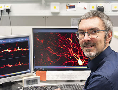
High-resolution microscopy images provide unique insights into brain cells and may contribute to a better understanding of learning and memory processes in the future: Marcel Lauterbach, Junior Professor of Molecular Imaging, was able to visualize almost all dendritic spines of a nerve cell in detail at the same time. He was able to show that these fine protrusions do not grow in the same way on every cell protrusion, nor are they distributed randomly. Instead, he discovered different arrangements of the five types of dendritic spines. The researcher published his findings in the journal “Progress in Neurobiology”. (Foto: © Laura Glücklich)
For further information and a link to the publication, please visit the UdS press release of February 1, 2024.
Hochauflösende Mikroskopie-Aufnahmen ermöglichen einzigartige Einblicke in Hirnzellen und können künftig zum besseren Verständnis von Lern- und Erinnerungsprozessen beitragen: Marcel Lauterbach, Juniorprofessor für Molekulare Bildgebung, konnte erstmals fast alle dendritischen Dornen einer Nervenzelle gleichzeitig detailliert sichtbar machen. Er konnte nachweisen, dass diese feinen Ausstülpungen nicht auf jedem Zellausläufer gleich wachsen und auch nicht zufällig auf ihnen verteilt sind. Vielmehr stellte er verschiedene Anordnungen der fünf Typen dendritischer Dornen fest.
Die Ergebnisse veröffentlicht der Forscher in der Fachzeitschrift “Progress in Neurobiology”. (Foto: © Laura Glücklich)
Weitere Informationen und einen Link zur Publikation finden Sie in der UdS-Pressemitteilung vom 2. Februar 2024.